
- All
- Product Name
- Product Keyword
- Product Model
- Product Summary
- Product Description
- Multi Field Search
Views: 0 Author: Site Editor Publish Time: 2026-10-08 Origin: Site


Cone beam computed tomography (CBCT) provides three-dimensional images of dental and craniofacial structures. In orthodontics, it can help clinicians evaluate complex cases when conventional two-dimensional radiographs do not provide enough diagnostic information.
Traditional panoramic and cephalometric images remain useful for many orthodontic assessments. However, overlapping anatomical structures can make it difficult to determine the exact position of impacted teeth, assess certain root conditions, or evaluate bone boundaries in three dimensions. CBCT offers multiplanar views that can support more detailed assessment in selected cases.
CBCT should not be considered a routine replacement for conventional imaging. Clinicians must balance its diagnostic value against radiation exposure, patient-specific needs, and available alternatives. This guide explains the main applications of CBCT in orthodontics, its clinical benefits, and the factors to consider before adopting it into a digital workflow.
Table of Contents
Key Takeaways
Three-dimensional assessment: CBCT helps evaluate impacted teeth, root relationships, skeletal structures, and selected bone conditions.
Case-specific use: CBCT is most useful when the additional information may affect diagnosis or treatment planning.
Radiation protection: Clinicians should justify each scan and use an appropriate field of view and exposure protocol.
Digital integration: Combining CBCT data with intraoral scans can support comprehensive orthodontic treatment planning.
Practice readiness: Software compatibility, staff training, image review protocols, and total ownership costs matter when implementing CBCT.
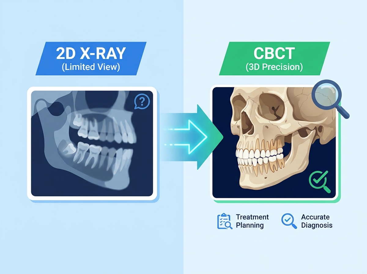
Understanding the differences between conventional radiography and CBCT helps clinicians decide when three-dimensional imaging may be appropriate.
Panoramic and cephalometric radiographs project three-dimensional anatomy onto a two-dimensional image. This makes them useful for many routine assessments, but some anatomical details may overlap or appear distorted.
Anatomical superimposition: Overlapping structures can make it difficult to assess the exact position of impacted teeth or their relationship with adjacent roots.
Magnification and distortion: Image geometry and patient positioning can affect measurements and interpretation.
Limited transverse assessment: Two-dimensional images provide limited information about buccal and lingual bone boundaries.
Restricted spatial information: A single projection cannot show the full three-dimensional relationship between teeth, roots, and surrounding structures.
CBCT acquires multiple projections and reconstructs them into a three-dimensional image volume. Clinicians can examine the anatomy in axial, coronal, and sagittal views, as well as in reconstructed views generated by suitable software.
CBCT may help clinicians:
Assess the position and orientation of impacted teeth.
Examine the relationship between roots and surrounding anatomical structures.
Evaluate selected bone dimensions and skeletal asymmetries.
Review craniofacial anatomy when planning complex orthodontic or surgical treatment.
CBCT measurements are not free from error. Image quality, voxel size, patient movement, artifacts, and the measurement method can all affect interpretation. The appropriate imaging method depends on the diagnostic question.
Imaging Feature | Traditional 2D Radiography | CBCT Imaging |
|---|---|---|
Anatomical view | Two-dimensional projection | Three-dimensional image volume |
Structure overlap | May obscure relevant anatomy | Reduces superimposition through multiplanar views |
Root and tooth localization | Limited by projection | Supports three-dimensional localization |
Bone assessment | Limited in some dimensions | Provides cross-sectional views of selected bone structures |
Typical role | Routine assessment when appropriate | Supplemental imaging for selected clinical questions |
CBCT can be valuable when a three-dimensional view may change diagnosis, treatment selection, or the management of a complex case.
Impacted maxillary canines and other ectopic teeth may be difficult to localize using conventional images alone. CBCT can help clinicians determine whether an impacted tooth lies buccally, palatally, or close to adjacent roots.
It may also help assess root morphology, the eruption path, and possible root resorption involving neighboring teeth. This information can support decisions about surgical exposure and orthodontic traction.
The scan should be prescribed when the additional information is expected to influence management. The smallest suitable field of view should be considered.
Three-dimensional imaging can support the assessment of complex skeletal discrepancies and craniofacial asymmetry. Clinicians can examine the relative positions and shapes of the maxilla, mandible, and other relevant structures.
In selected cases, CBCT may contribute to planning palatal expansion or orthognathic surgery. Assessment of midpalatal suture maturation and temporomandibular joint structures should be based on the specific clinical indication and the limitations of the available evidence.
CBCT can display upper airway morphology and estimate dimensions within the scanned region. This may provide supplementary anatomical information when airway structures are relevant to a patient's assessment.
However, airway volume or a single cross-sectional measurement cannot independently diagnose obstructive sleep apnea or determine airway function during sleep. Suspected sleep-related breathing disorders require appropriate clinical assessment and, when indicated, sleep testing.
The position of teeth relative to the surrounding alveolar bone can be important when planning certain orthodontic movements. In selected cases, CBCT cross-sectional images may help evaluate buccal and lingual bone boundaries or suspected defects that are difficult to assess with conventional imaging.
These findings should be interpreted alongside clinical periodontal examination and other relevant records. CBCT should not be used routinely for every patient solely to measure bone thickness.
CBCT data can support three-dimensional analysis of craniofacial structures in selected complex cases. Suitable software allows clinicians to orient the image volume, review anatomical landmarks, and assess skeletal relationships from multiple perspectives.
For orthognathic surgery planning, CBCT may also be integrated with other digital records to help visualize skeletal relationships. The accuracy of any simulation depends on the quality of the source data, the software, and the clinical planning process.
The main benefit of CBCT is access to additional anatomical information when conventional imaging does not adequately answer a clinical question. Its value depends on whether that information can improve decision-making.
Three-dimensional views can help clinicians understand the position of impacted teeth, the relationship between roots and surrounding structures, and selected skeletal conditions. This information may support decisions about treatment mechanics, surgical coordination, or referral.
CBCT does not guarantee shorter treatment times or eliminate complications. Treatment outcomes still depend on diagnosis, clinical judgment, patient factors, and ongoing monitoring.
Three-dimensional images can help clinicians explain selected anatomical findings to patients. For example, a reconstructed view may make the position of an impacted tooth easier to understand than a two-dimensional projection alone.
Visual explanations can support informed discussions about treatment options, benefits, limitations, and risks. They should complement, rather than replace, a balanced clinical explanation.
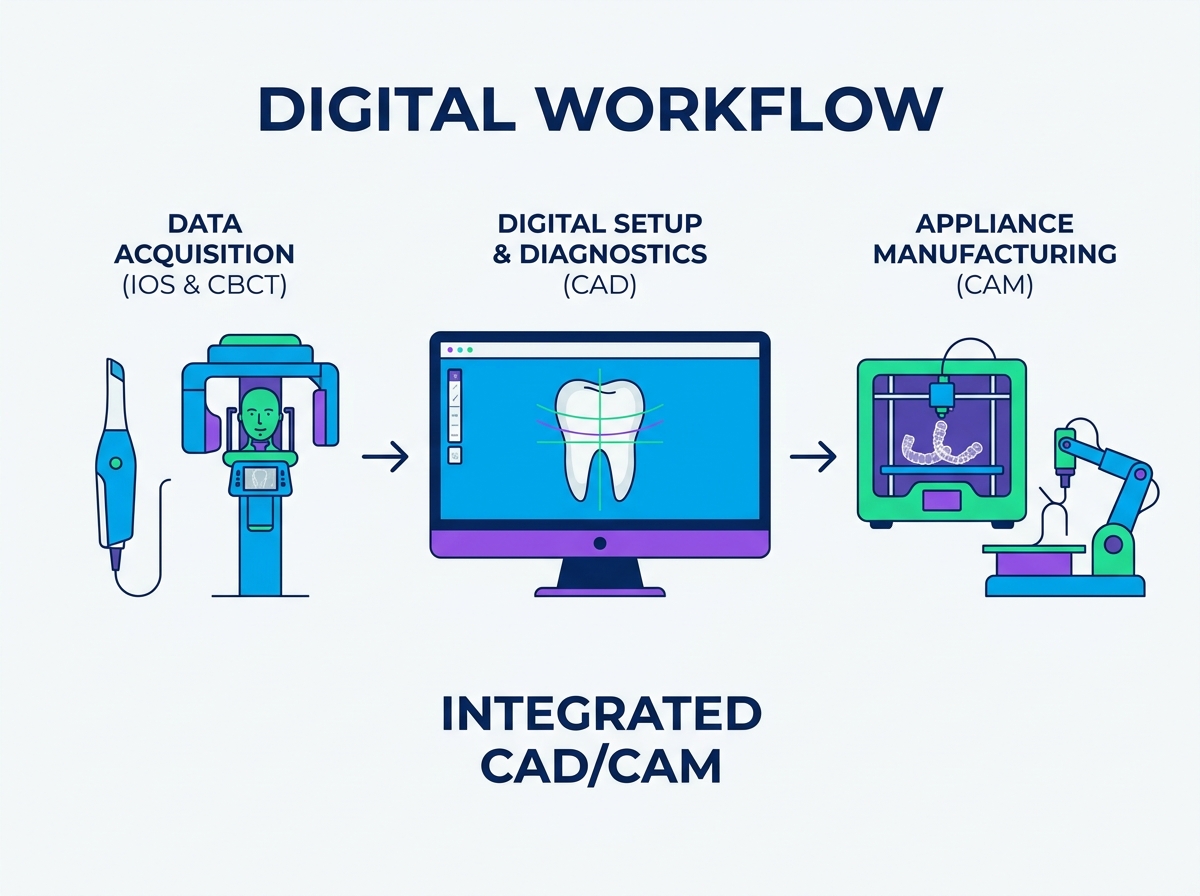
CBCT data may be combined with intraoral surface scans to provide complementary information. Intraoral scans capture visible tooth and gingival surfaces, while CBCT can show roots and selected underlying structures.
When the software supports reliable data registration, clinicians can review these datasets together during treatment planning. Learn more about digital orthodontics and integrated workflows.

For clear aligner workflows, CBCT integration may provide additional root and bone information when clinically justified. Explore SmileyNova's orthodontic treatment planning service for an example of a digital planning workflow that incorporates CBCT data.
CBCT should be prescribed when the expected diagnostic benefit justifies the radiation exposure and the information is likely to influence patient management. It should not be used automatically for every new orthodontic patient.
Radiation protection is central to responsible CBCT use. ALARA means “As Low As Reasonably Achievable,” while ALADA emphasizes keeping exposure “As Low As Diagnostically Acceptable.”
Clinicians should:
Confirm the clinical question before requesting a scan.
Consider whether existing records or lower-dose imaging can answer the question.
Select exposure settings appropriate for the patient and diagnostic task.
Use the smallest field of view that adequately covers the region of interest.
Avoid repeat scans unless additional imaging is clinically justified.
Ensure that the scanned volume is appropriately reviewed and documented.
The appropriate protocol depends on the patient, the device, and the intended assessment. Pediatric and adolescent patients require particular attention to justification and dose optimization.
The field of view (FOV) determines how much anatomy is included in the scan. A larger FOV may capture more structures, but it can also expose more tissue than necessary.
FOV Consideration | Potential Use | Key Consideration |
|---|---|---|
Small FOV | A localized issue, such as an impacted tooth | Include enough anatomy to answer the diagnostic question |
Medium FOV | Selected assessments involving multiple teeth or broader jaw regions | Confirm that the scan range is clinically necessary |
Large FOV | Selected craniofacial or orthognathic surgery assessments | Justify the wider scan range and review the full volume |
These categories are illustrative, not universal size standards. Actual FOV dimensions and available protocols vary by device.
Depending on the individual case, CBCT may be considered for:
Impacted teeth when localization or suspected adjacent root resorption may affect treatment.
Complex craniofacial conditions or significant skeletal asymmetry.
Selected cases requiring three-dimensional surgical planning.
Specific questions about root position or alveolar bone boundaries that cannot be answered adequately by other methods.
Other complex diagnostic situations where three-dimensional information is expected to change management.
The indication should be documented, and clinicians should follow applicable professional guidance and local regulations.
Adopting CBCT involves more than purchasing imaging equipment. Practices should evaluate workflow needs, data management, staff competency, and responsibilities for image interpretation.
Before choosing a CBCT system, confirm that its data can be exported and used by the practice's existing software. DICOM is commonly used for medical imaging data, while intraoral surface scans are often supplied in formats such as STL or PLY.
If the workflow requires merging CBCT and intraoral scan data, check whether the software supports the required registration process and whether the team can verify the alignment. Learn more about clear aligner treatment planning.
Staff should be trained in patient positioning, scan preparation, image quality checks, and the practice's referral or review procedures. Clinicians also need sufficient training to navigate multiplanar views and recognize the limitations of the images.
Standardized protocols can reduce avoidable errors and help teams identify when a scan may need further review.
CBCT may include anatomical areas beyond the immediate orthodontic concern. Unexpected findings may involve the sinuses, jaws, or other structures within the scanned volume.
Practices should establish a clear process for reviewing the full image volume, documenting relevant findings, and arranging referral or specialist interpretation when needed. Responsibilities vary by jurisdiction and professional role, so local requirements should be followed.
A practice considering CBCT should assess the total cost of ownership rather than focusing only on the purchase price. Relevant factors include installation, maintenance, software licensing, staff training, image storage, and any specialist reporting services.
Potential operational benefits should be evaluated against the practice's actual case mix and referral needs. CBCT is not automatically cost-effective for every clinic, and clinical justification should remain the primary consideration.
CBCT in orthodontics provides three-dimensional anatomical information that can support diagnosis and treatment planning in selected complex cases. It is particularly relevant when conventional imaging cannot adequately show the position of impacted teeth, root relationships, or specific skeletal structures.
However, CBCT is not a routine replacement for panoramic or cephalometric radiography. Clinicians should justify each scan, optimize radiation exposure, select an appropriate field of view, and ensure that the full scanned volume is reviewed.
For practices integrating CBCT into digital orthodontics, software compatibility, staff training, data management, and clear clinical protocols are essential. The goal is not to scan every patient, but to obtain the right information when it can make a meaningful difference to patient care.
CBCT provides three-dimensional views of dental and craniofacial structures. In selected cases, it can help assess impacted teeth, root relationships, skeletal conditions, and other anatomical questions that conventional imaging cannot adequately answer.
No. CBCT should be prescribed when the expected diagnostic benefit justifies the radiation exposure. Many routine orthodontic cases can be assessed using clinical examination and appropriate conventional imaging.
CBCT provides three-dimensional views and reduces anatomical superimposition. However, image quality and measurement accuracy depend on scan settings, artifacts, patient movement, and the specific diagnostic task. It is not automatically the best choice for every clinical question.
No. CBCT can show airway anatomy and estimated dimensions, but it cannot independently diagnose obstructive sleep apnea. Diagnosis requires appropriate clinical assessment and, when indicated, sleep testing.
The field of view should be large enough to answer the clinical question but no larger than necessary. Clinicians should consider the anatomical region, patient characteristics, device capabilities, and radiation protection principles when selecting the scan protocol.


