
- All
- Product Name
- Product Keyword
- Product Model
- Product Summary
- Product Description
- Multi Field Search
Views: 0 Author: Site Editor Publish Time: 2026-09-18 Origin: Site


Orthodontics historically relied on reactive, trial-and-error adjustments. Clinicians applied forces, waited for biological responses, and corrected course during subsequent visits. This methodology required constant monitoring and frequent physical interventions. Today, patients and practitioners demand precision-driven, predictable clinical workflows. Unpredictable force systems carry severe compounding costs for any modern clinic. Prolonged therapy periods frustrate patients and delay case completion. Unexpected mid-course corrections consume valuable chair time. Poorly planned forces compromise root integrity and lead to severe clinical complications. Patient dissatisfaction ultimately erodes practice reputation and operational efficiency.
To solve these clinical problems, modern practices adopt advanced computational tools. We use tooth movement simulation as the bridge between theoretical biomechanics and clinical reality. This technology enables practitioners to validate treatment strategies computationally before applying physical forces. By modeling the exact biological and mechanical constraints of each patient, you eliminate clinical guesswork and ensure physical execution matches clinical intent.
Predictability Over Guesswork: Advanced simulation shifts treatment planning from aesthetic estimations to biomechanically sound, mathematically verified force applications.
Methodological Evolution: The integration of the Finite Element Method (FEM) and AI-driven spatial analysis allows for accurate modeling of complex contact boundary conditions and long-term movement.
Biological Constraints: High-tier simulation tools differentiate themselves by accounting for actual physiological responses, such as bone remodeling, rather than just geometric morphing.
Practice ROI: Implementing robust simulation software directly correlates with reduced chair time, fewer refinement stages, and higher case acceptance rates through transparent patient reporting.
Traditional cephalometric and panoramic assessments contain inherent clinical blind spots. They compress complex 3D spatial movements into flat, two-dimensional planes. You cannot accurately predict torquing, tipping, or rotational forces using 2D imaging alone. The transverse dimension is almost entirely lost in standard lateral cephalograms, leaving clinicians to guess the true buccolingual inclination of posterior segments.
Manual force estimation often fails to account for complex reciprocal forces. Newton's third law dictates that every applied force generates an equal and opposite reaction. When you retract anterior teeth, the posterior anchorage segment experiences a mesializing force. Traditional planning struggles to map dynamic force decay over extended periods and routinely misses unwanted anchorage loss during space closure. This leaves clinicians reacting to unexpected shifts rather than preventing them proactively. Relying on 2D data for 3D problems guarantees clinical inefficiency.
Diagnostic Variable | Traditional 2D Planning | 3D Biomechanical Simulation |
|---|---|---|
Root Position | Estimated based on average crown-root angles | Accurately mapped using CBCT DICOM data |
Anchorage Control | Reactive monitoring during appointments | Proactive calculation of reciprocal forces |
Transverse Dimension | Blind spot in lateral cephalograms | Full 360-degree spatial awareness |
Force Decay | Assumed based on generalized material properties | Calculated dynamically over the treatment timeline |
Basic aesthetic aligner setups often rely entirely on geometric morphing. They simply interpolate the shortest path between point A and point B on a computer screen. This visual trickery is not true biomechanical movement. Teeth do not float freely in empty space. They sit firmly anchored in the periodontal ligament (PDL) and alveolar bone. Every tooth has a specific center of resistance, typically located one-third to one-half the distance down the root embedded in bone. Geometric morphing ignores this center of resistance, applying virtual forces to the crown without calculating the resulting moments that cause unwanted tipping.
The physiological reality involves active, continuous bone remodeling. Failing to simulate this biological resistance leads directly to clinical surprises. When software ignores the biological envelope, you encounter severe complications:
Cortical Plate Collisions: Roots get pushed against or through the dense cortical bone, causing fenestrations or dehiscences.
Root Resorption: Excessive, concentrated forces on the root apex lead to permanent blunting and loss of tooth structure.
Tracking Failures: The physical tooth stops moving because the biological resistance exceeds the applied mechanical force, while the plastic aligner continues to advance.
Periodontal Compromise: Unplanned expansion pushes teeth outside the bony housing, leading to severe gingival recession.
The Finite Element Method (FEM) serves as the industry standard for numeric simulation models in orthodontics. FEM breaks down complex continuous structures into smaller, calculable elements called nodes. Software models the periodontal ligament, alveolar bone, and individual tooth structures as a highly detailed 3D mesh. By applying mathematical equations to these nodes, the system predicts stress and strain distribution accurately.
Biological tissues possess non-linear, viscoelastic properties. The PDL acts as a shock absorber, distributing forces differently depending on the load's magnitude and direction. FEM calculates exactly how these tissues will deform under specific mechanical loads. This transforms a visual guess into a physics-based certainty. FEM allows clinicians to see the invisible forces acting beneath the gingival margin. It provides a mathematically verified roadmap for safe, effective orthodontic therapy.
Machine learning algorithms now analyze vast datasets of historical case data. They predict clinical tooth movement trajectories based on thousands of documented outcomes. This helps minimize surprises during treatment by flagging high-risk movements early. For example, AI can identify that a specific extrusion movement on a lateral incisor has a high probability of failure based on historical tracking data.
However, pure pattern recognition is never enough for clinical safety. The best systems balance AI spatial analysis with physics-based mathematical calculations in 3D space. AI provides a clear picture of likely movement based on past successes. The physics engine ensures that this proposed movement obeys the strict laws of biology and mechanics. This hybrid approach delivers both speed and clinical safety. It removes human error from initial staging while enforcing strict biomechanical boundaries.
Simulation software computes highly complex force-moment systems instantly. It tracks how these forces dynamically change throughout the entire therapy period. As teeth move, the applied force decays or shifts direction entirely. A power chain loses elasticity. An aligner plastic undergoes stress relaxation. The software recalculates these vectors continuously to maintain accuracy.
It also models critical contact boundary conditions that dictate real-world outcomes. Factoring in these dynamic boundaries is mandatory for predicting long-term orthodontic tooth movement accurately. Key boundary conditions include:
Tooth-to-Tooth Collisions: Preventing crowns or roots from intersecting during space closure or crowding resolution.
Bracket-Wire Interactions: Calculating the friction and play between a specific wire alloy and the bracket slot.
Occlusal Interferences: Identifying premature contacts during the transition phases of treatment that could dislodge appliances or cause traumatic occlusion.
Attachment Mechanics: Evaluating how the geometry of a composite attachment alters the force vector delivered by an aligner.
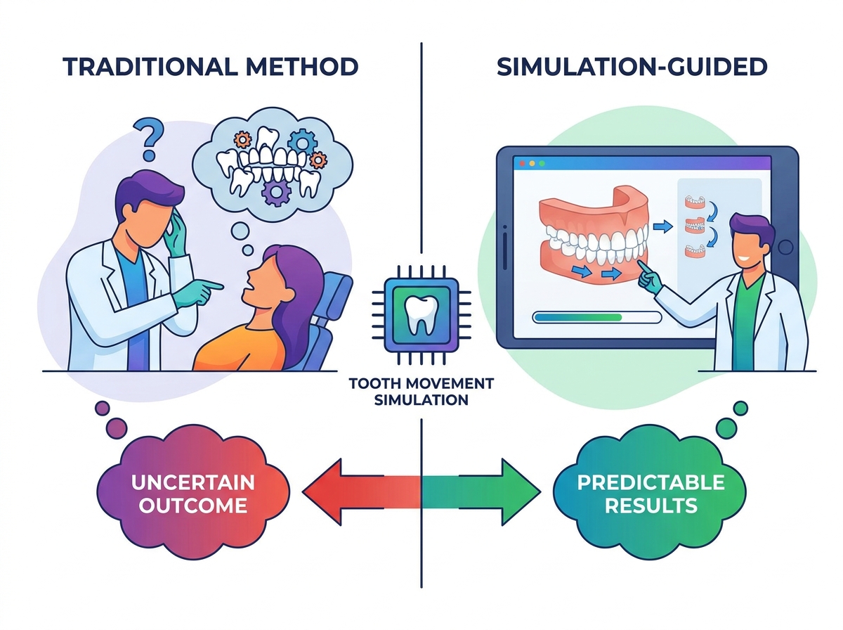
You must evaluate whether a software platform respects actual biological limits. The system must prevent roots from moving outside the cortical plate computationally. If the software allows impossible movements, it is useless for clinical planning. You need tools that enforce hard stops when proposed forces exceed physiological tolerances. This protects the patient from iatrogenic damage.
Use this checklist when evaluating simulation platforms for your clinic:
Does the software allow for custom bone density inputs based on patient age and Hounsfield units from a CBCT?
Can you integrate true root morphology directly via DICOM files rather than relying on generic library roots?
Does the system provide an accurate, dynamic representation of the center of resistance for each tooth as it moves through bone?
Are periodontal ligament thickness variations accounted for in the physics engine?
Does the platform flag collision risks between adjacent roots automatically during staging?
Seamless integration with your existing clinic infrastructure is non-negotiable. The software must ingest data from intraoral scanners, CBCT machines, and CAD/CAM systems without data loss. Beware of proprietary lock-in when selecting a vendor. Closed ecosystems force you to buy specific hardware and restrict your manufacturing options.
Open-architecture platforms support standard STL, PLY, and DICOM workflows natively. This flexibility protects your existing hardware investments. It also keeps your lab options open for manufacturing appliances. You should control your clinical data entirely. Open systems allow you to route files to in-house 3D printers or external fabrication partners freely, giving you total control over your supply chain.
Comprehensive orthodontic treatment programs generate dual-purpose outputs. First, they produce highly technical clinical reports for lab technicians and treating doctors. These reports detail anchorage requirements, IPR amounts, and staging sequences. Second, they create automated, visual reports designed specifically for users and patients.
Visual simulations translate complex biomechanics into digestible, high-conversion visual aids. Patients do not understand force-moment vectors or anchorage values. They understand a clear, 3D time-lapse of their own treatment journey. Showing them a biologically accurate simulation builds immediate trust. It dramatically increases case acceptance rates for high-value treatments. Transparent communication tools turn a clinical necessity into a powerful practice growth engine.
Unexpected clinical surprises carry a heavy operational cost for your practice. Extra visits consume valuable chair time that could go to new consultations. New intraoral scans disrupt the daily schedule and frustrate clinical staff. Additional lab fees eat directly into your profit margins on every case. When a patient requires three rounds of refinements, the profitability of that case drops to near zero.
Accurate simulation reduces the overall therapy period drastically. By getting the force system right the first time, you eliminate unnecessary refinement stages. Predictable tracking means fewer emergency visits for off-track appliances or poking wires. This results in a much higher hourly production rate for the clinician. You complete cases faster, increase patient turnover, and maximize clinic profitability.
Metric | Without Simulation (Trial & Error) | With Biomechanical Simulation |
|---|---|---|
Average Refinements | 2 to 4 per case | 0 to 1 per case |
Chair Time per Patient | High (Frequent troubleshooting) | Low (Scheduled monitoring only) |
Treatment Duration | 18 to 24 months | 12 to 16 months |
Material/Lab Costs | High (Multiple appliance orders) | Optimized (Single comprehensive order) |
Simulation allows clinicians to computationally test multiple treatment strategies before committing to a clinical path. You can run side-by-side comparisons of extraction versus non-extraction approaches safely. You can evaluate interproximal reduction (IPR) against arch expansion to see which yields better functional occlusion and facial aesthetics.
This empowers practitioners to confidently take on complex interdisciplinary cases. Surgical orthodontics and severe impactions no longer require blind faith in basic setups. You can map the entire biological response beforehand. Cases you might have previously referred out stay in-house. This drives significant practice growth and establishes your clinic as a destination for advanced, predictable care.
There is a severe clinical danger in trusting automated AI simulations blindly. Software is a decision-support tool, not a replacement for clinical expertise and diagnostic judgment. If you do not apply professional biomechanical oversight, you risk executing fundamentally flawed treatment plans.
The "black box" problem occurs when a clinician accepts an AI-generated outcome without understanding the underlying force mechanics. Algorithms optimize for visual alignment, sometimes at the expense of periodontal health. Always verify the simulation against your own diagnostic judgment. You must review the force vectors, check the anchorage requirements, and ensure the proposed movements respect the patient's unique biology. A beautiful digital setup means nothing if it pushes the lower incisors entirely out of the symphysis.
Adopting 3D tooth movement analysis software requires a realistic onboarding timeline. Staff and clinicians need time to adjust to new digital workflows and interfaces. Expect a learning curve of three to six months before the process feels entirely seamless. Rushing the implementation phase often leads to staff frustration and abandoned technology.
To mitigate disruption and ensure a smooth transition, implement these strategies:
Start with straightforward, single-arch cases to build initial team confidence.
Utilize all available vendor training sessions for your clinical and administrative staff.
Conduct parallel planning during the transition phase, comparing the digital simulation with your traditional setup methods.
Assign a dedicated digital workflow champion within your staff to manage file routing and basic troubleshooting.
Establish clear protocols for merging CBCT and intraoral scan data before uploading to the simulation platform.
Advanced simulation is no longer an optional luxury for elite practices. It is a baseline requirement for delivering evidence-based, predictable orthodontic therapy. Buyers must prioritize software that combines FEM-based biomechanical accuracy with seamless CBCT and intraoral scanner interoperability. Avoid tools that only offer superficial geometric morphing, as they fail to account for biological reality.
Take these immediate next steps to modernize your clinical workflow:
Audit your current refinement rates to identify the true financial cost of unpredictable tracking in your practice.
Request technical software demos focusing specifically on complex boundary condition handling and root collision detection.
Run a trial case utilizing your own patient CBCT data to verify the software's biological constraint accuracy.
Train your treatment coordinators to use 3D visual reports during patient consultations to boost case acceptance.
A: It is a computational method using physics, mathematics, and 3D modeling to predict how teeth will respond to specific orthodontic forces over a therapy period. It replaces guesswork with verified biomechanical planning.
A: Accuracy depends entirely on the underlying technology. FEM and CBCT-integrated models are highly accurate because they map real anatomy. Conversely, purely visual AI models often lack biological constraint awareness, reducing their clinical reliability.
A: FEM calculates the exact stress and strain on the periodontal ligament and alveolar bone. This allows the software to predict realistic biological responses to applied forces, ensuring proposed movements are physically possible.
A: AI is excellent for initial staging and pattern recognition to provide a clear picture of likely movement. However, it must be paired with strict biomechanical rules to prevent clinically impossible outcomes and iatrogenic damage.
A: Advanced numeric models factor in the physiological process of bone resorption on the pressure side and deposition on the tension side. This ensures the digital plan respects the natural speed and limits of human biology.
A: Aesthetic setups visually interpolate the shortest path between current and final tooth positions. Biomechanical simulation calculates the actual force required, dynamic boundary conditions, and the biological path of least resistance for safe movement.


